
Fluorescent in situ hybridisation
Fluorescent in situ hybridisation
Fluorescent in situ hybridisation (FISH) is a technique that, using fluorescent labelled DNA probes (often derived from fragments of DNA that were isolated during the HGP), can detect and confirm gene and chromosome abnormalities beyond the resolution of routine cytogenetics. OR
Fluorescence in situ hybridization (FISH) provides researchers with a way to visualize and map the genetic material in an individual’s cells, including specific genes or portions of genes. This is important for understanding a variety of chromosomal abnormalities and other genetic mutations. Unlike most other techniques used to study chromosomes, FISH does not have to be performed on cells that are actively dividing. This makes it a very versatile procedure.
Sample DNA is first denatured, converting double-stranded to single-stranded DNA. The fluorescent labelled probe (complementary to the DNA sequence of interest) is then added to the single-stranded DNA. If the DNA sequence of interest is present in the sample, the probe hybridises with the complementary bases as the DNA re-forms back into a double helix.
The probe signal can then be detected through a fluorescence microscope and the sample DNA scored for the presence or absence of the signal.

FISH can be performed using two sample types: metaphase chromosomes and interphase nuclei.
Metaphase FISH
FISH can be performed on metaphase chromosomes to detect specific micro-deletions undetectable by routine techniques, or to identify chromosome translocations or extra material of unknown origin.
Microdeletion syndromes currently detectable using FISH
- Cri-du-chat syndrome:
- Results from the deletion of part of the short arm of chromosome 5.
- The main clinical feature is the presence of a high-pitched ‘cat-like’ cry present in the newborn that may
disappear with age. Other features include a round, full face, widely spread eyes (hypertelorism), an extra fold of skin at the inner corners of the eyes (epicanthal folds), a flattened and widened nasal bridge and ears that are positioned low on the head, severe cognitive, speech and motor delays, and feeding problems from birth which may lead to poor growth
- Miller–Dieker syndrome:
- Results from the deletion of several adjacent genes in the short arm of chromosome 17 (17 p).
- Clinical features include lissencephaly and a characteristic facial appearance (prominent forehead with bitemporal hollowing, short nose with upturned nares, thickened upper lip with a thin vermilion upper border, widely spaced eyes, low ears, and small jaw). The syndrome may result in mental retardation, epilepsy, preand postnatal growth retardation, and reduced lifespan. There may also be multiple
abnormalities of the brain, kidneys, heart, and gastrointestinal tract.
- Smith–Magenis syndrome:
- Results from a microdeletion in the short arm of chromosome 17 [del(17)(p11.2 p11.2)].
- As well as characteristic facial abnormalities (short flat head, prominent forehead, broad square
face, upslanting eyeslits, deep-set eyes, underdeveloped midface, broad nasal bridge, short
nose and tented upper lip), the syndrome may also cause mild-to-moderate mental retardation.
- Steroid sulphatase deficiency (Also known as X-linked ichthyosis)
- It is a genetic disorder of the skin that occurs only in males.
- The condition develops in infancy and manifests as tan or grey scales on the skin that are a result of a deficiency in the enzyme steroid sulphatase due to genetic mutations of the gene.
- DiGeorge syndrome (also known as velocardiofacial/CATCH-22/Shprintzen syndrome)
- Kallman syndrome
- Williams syndrome
- Wolf–Hirschhorn syndrome:
- Results from a partial deletion of the short arm of chromosome 4.
- Many parts of the body are affected by this syndrome as the deletion affects fetal growth and development. Common features include profound learning disability, microcephaly, seizures, low muscle tone and poor muscle development, heart defects, and cleft lip and/or palate
Interphase FISH
FISH can be used in interphase cells to determine the chromosome number of one or more chromosomes as well as to detect some specific chromosome rearrangements characteristic of certain cancers. The advantage of interphase FISH is that it can be performed very rapidly as cell growth is not required.
An example of interphase FISH is the aneuploid screen test performed on amniotic fluid cells to determine the presence of the common trisomies. Sample nuclei are denatured and incubated with probes for chromosomes 13, 18, 21, X and Y.
Comparison of FISH with other techniques of gene mapping
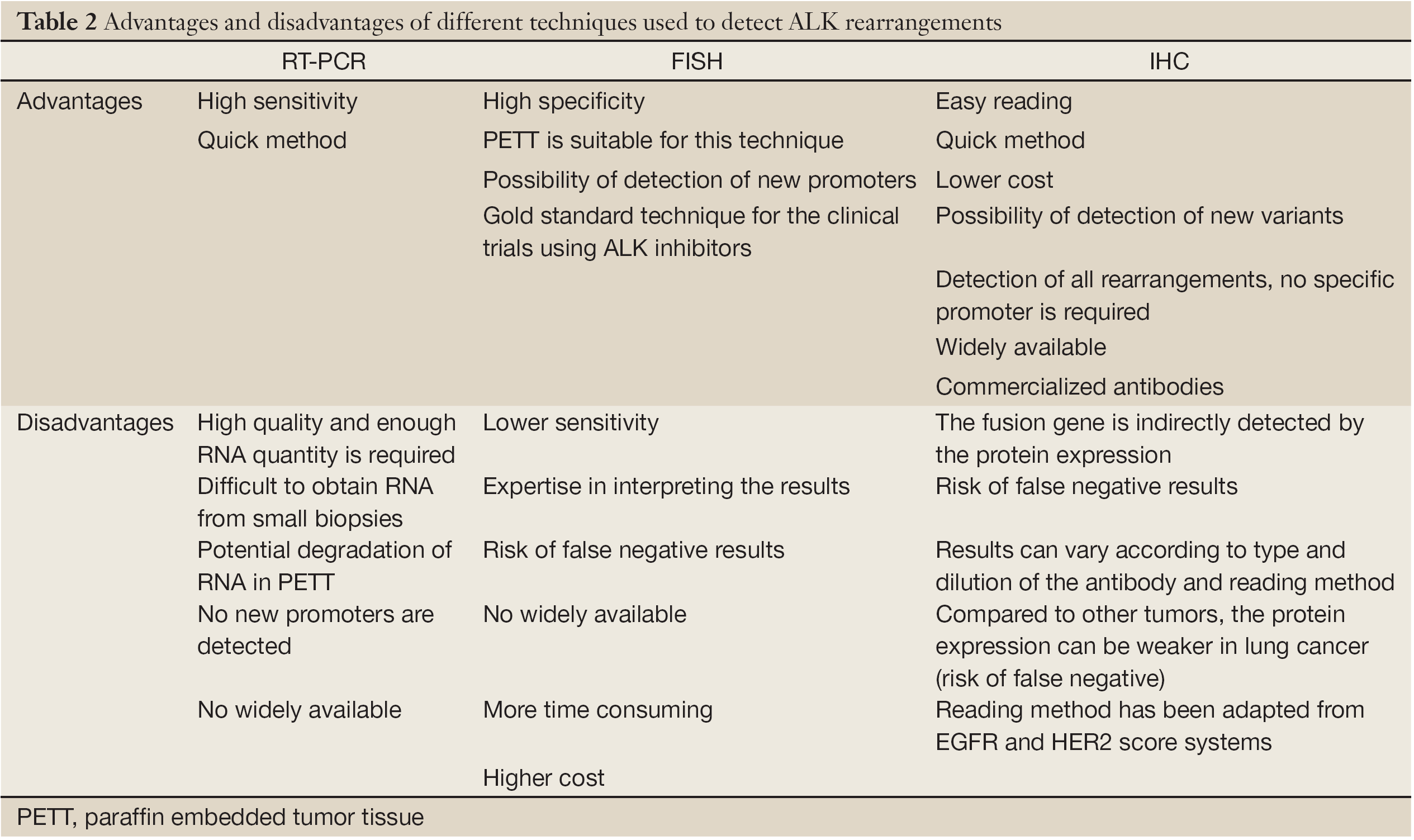
Image curtsy: Nature,